Monkeypox Symptoms and Signs
The incubation period for monkeypox, from the time of infection to the onset of disease symptoms, is usually 6–14 days, but can last 5–21 days, depending on the degree of invasiveness of contact with an infected host. [1] [2]
According to seroepidemiological studies in Africa, most people are asymptomatic with monkeypox infection. In the case of symptomatic disease, however, it manifests systemically as fever, headache, chills, exhaustion, asthenia, back pain, and muscle aches. The clinical picture can vary depending on the strain of monkeypox virus. [3]
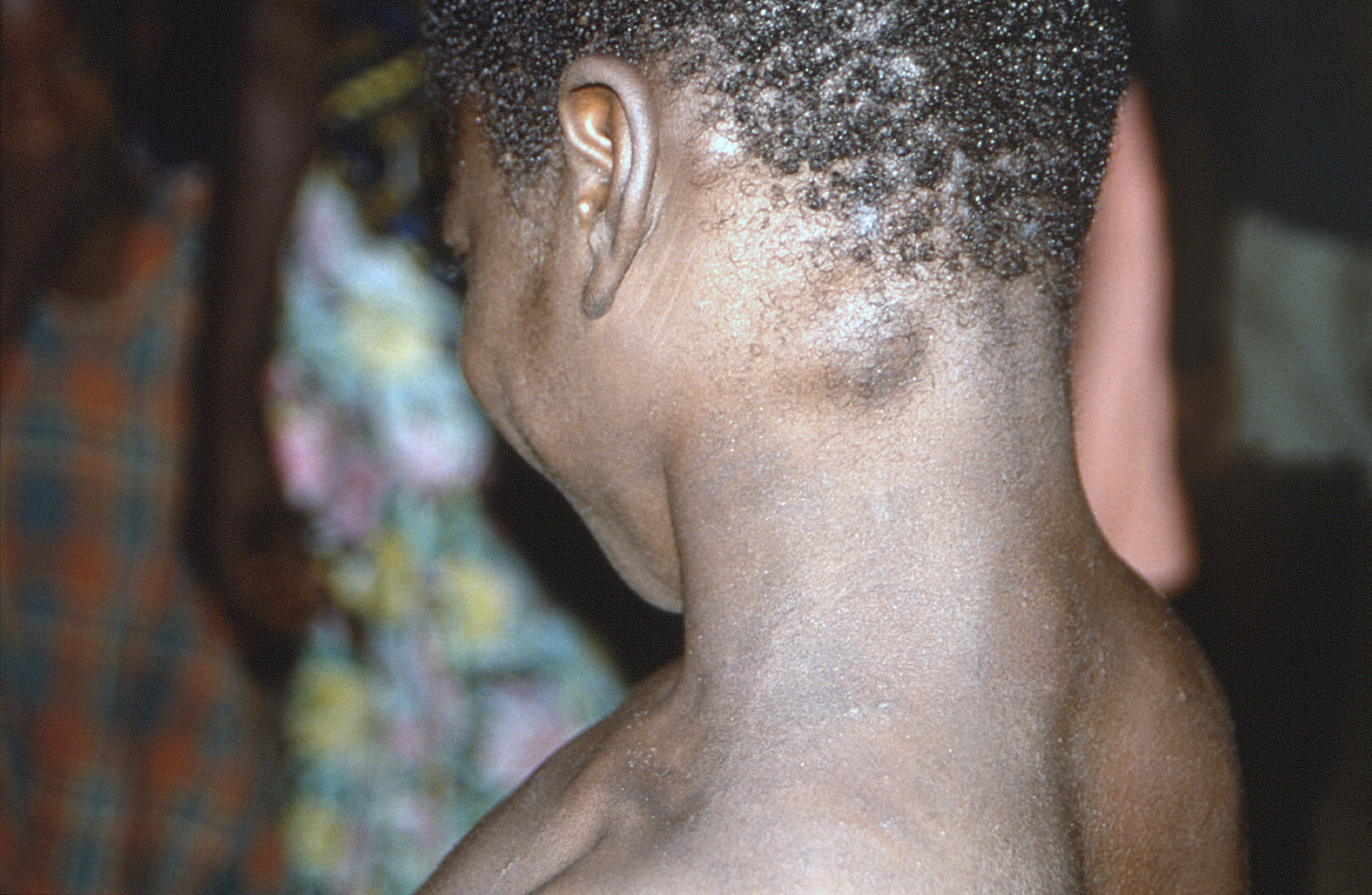
Lymphadenopathy (enlarged lymph nodes) is a characteristic feature of monkeypox, making it possible to distinguish it from other infections presenting in a similar way, such as chickenpox (varicella), measles, smallpox. Lymphadenopathy may occur in the submandibular, cervical, axillary, or inguinal regions. The enlarged nodes are firm, tender, and sometimes painful. The presence of lymphadenopathy indicates perhaps more effective immune recognition and immune response to monkeypox than in smallpox. [4] [5] [6]
Within 1 to 3 days (sometimes longer) of the onset of fever, a rash appears, usually affecting the face first and then spreading to other parts of the body.
The appearance of the rash is considered the beginning of the infectious period. [2] [7] [8] [9] Although, it is believed that persons with prodromal (pre-disease) symptoms can also transmit monkeypox virus. [2]
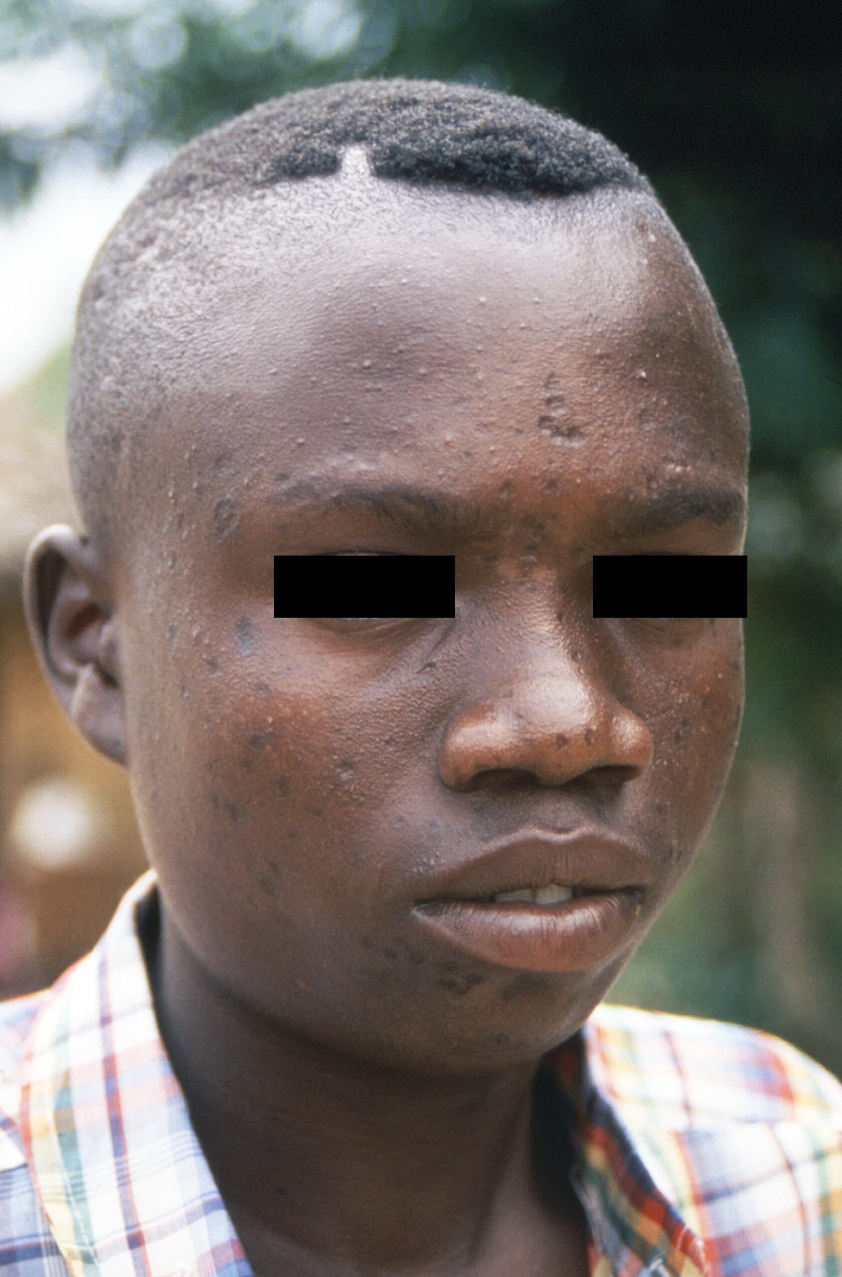
The rash in monkeypox is usually localized to the face (95% of cases) and to the extremities, the palms and soles of the feet (75%), rather than the torso. The size of lesions, which may also occur on the oral mucosa (70%), genitals (30%), conjunctiva (20%), and cornea, is 0.5–1 cm in diameter. [1]
The rash develops sequentially, going through several stages over a period of 1–2 weeks.
- First, an enanthema (a rash on the mucous membranes) appears on the tongue and mouth.
- Following this, the rash appears on the face as macules (lesions with a flat base), which then spread to the arms and legs and then to the hands and feet, including the palms and soles.
- Within 24 hours, the rash takes over all parts of the body with a centrifugal distribution (concentrated on the face, hands and feet).
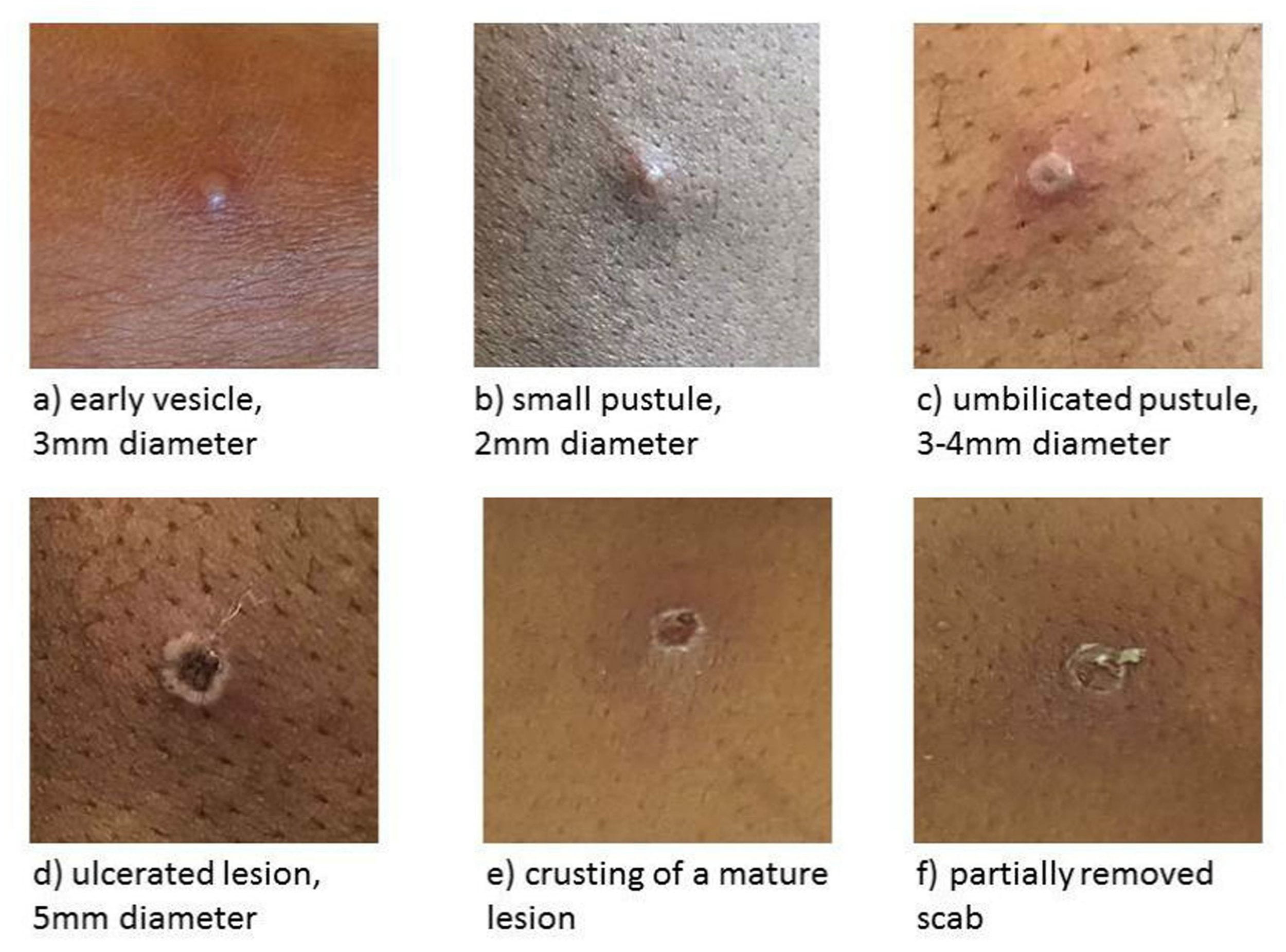
- Around day 3, papules (slightly raised and hard lesions) form.
- On day 4–5, the lesions turn into vesicles (raised and filled with clear fluid).
- By day 6–7, they transform into pustules (filled with opaque yellowish fluid), which are usually sharply elevated, round, deep-set, and firm to the touch; then there is umbilication of pustules (a depression forms in their center).
- The pustules persist for 5–7 days; by the end of 2 weeks, they dry out, becoming crusty and scabby, which remain for another 1 week before desquamation (peeling and falling off). [2] [5] [10]
After the scabs have fallen off, ulcerative scars and/or areas of lighter or darker skin may remain. Once all scabs have fallen off, the person is no longer contagious.
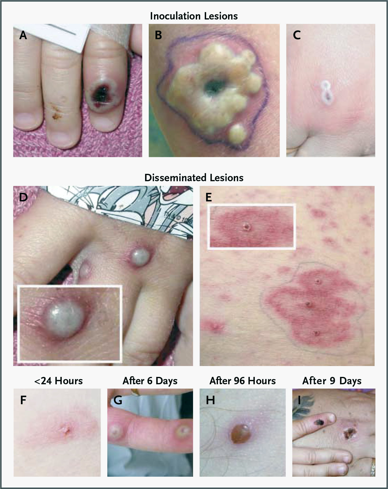
The number of lesions varies from single to several thousand, which in severe cases may coalesce up to the detachment of large areas of skin. Sometimes only a localized rash on the hands develops, due to direct contact with an infected host. [11]
The lesions can be very itchy, and if they are scratched, there is a risk of secondary bacterial infection.
Monkeypox usually lasts 2–4 weeks, eventually healing itself to full recovery.
The severity of monkeypox depends on the initial general health status of the patient (comorbidity), the route of infection (invasive or noninvasive), and the virus strain. The West African clade (genetic group) of monkeypox virus is associated with a lighter course of the disease, fewer deaths (case fatality rate up to 3.6%) and limited human-to-human transmission. Infection caused by monkeypox virus of the Congo Basin clade (Central African clade) is more severe, characterized by a higher mortality (case fatality rate up to 10.6%, death usually occurs in the 2nd week of illness) and increased human-to-human transmission. [12] [13] [14] [15]
Persons with a weakened immune system (very young children, the elderly, pregnant women, with immune deficiency) are at risk of a severe course of monkeypox with probable complications and death. [16]
Possible complications of monkeypox include respiratory failure, secondary bacterial infections, encephalitis, conjunctivitis, keratitis, and pneumonia. Complications, however, are mostly true in endemic countries because of the low level of medical care provided. [1] [17]
Among the possible sequelae of monkeypox are disfiguring scars and permanent corneal lesions. [10]
According to data collected during the 2003 monkeypox outbreak in the United States, the dominant signs and symptoms of infection were as follows: rash (in 97% of cases), fever (85%), chills (71%), lymphadenopathy (71%), headache (65%), myalgia (56%). The onset of fever preceded the onset of rash by approximately 2 days, and the mean duration of fever was shorter than that of rash (8 and 12 days, respectively). [12]
Some patients (n=9/34) were hospitalized for a variety of reasons, such as nausea, vomiting, and dysphagia. Numerous nonspecific abnormalities in laboratory values were noted, including increased aminotransferases, leukocytosis, mild thrombocytopenia, and hypoalbuminemia. At discharge, the two most severe patients were diagnosed with encephalopathy and retropharyngeal abscess.
All patients survived thanks to supportive therapy; antiviral treatment was not administered. These favorable outcomes are associated with a healthier patient population, greater availability of supportive care, and a less virulent strain of monkeypox, which was imported from West Africa’s Ghana.
Monkeypox Diagnosis
Diagnostic assays for monkeypox include virus isolation (in mammalian cell cultures), electron microscopy, real-time polymerase chain reaction (PCR), enzyme-linked immunosorbent assay (ELISA), immunofluorescent antibody assay (IFA), and immunohistochemistry (IHC). [1] [2]
Electron microscopy reveals characteristic brick-shaped poxvirus virions. Histopathological analysis shows ballooning degeneration of keratinocytes, marked spongiosis, dermal edema, and acute inflammation, although these signs can also be seen in other viral infections. [3]
The possibility of detecting the monkeypox virus DNA genome by real-time PCR is well established in several laboratories in Europe. Scrapes, swabs and aspirated fluid from the lesion are preferable to blood samples because of the limited duration of viremia. The results of these samples correlate best with both the infectivity and the clinical course of the infection. Modern real-time PCR methods allow not only to differentiate monkeypox virus from other orthopoxviruses, but also to distinguish between two clades of this virus.
Using sera from patients collected during the 2003 monkeypox outbreak in the United States, ELISA was developed to detect class M (IgM) and class G (IgG) immunoglobulins to monkeypox virus. Serum IgM and IgG antibodies were detected 5 and 8 days after rash onset, respectively. [4]
Serology has limited value because of immunological cross-reactivity between human pathogenic orthopoxviruses, although it can be useful in excluding recent orthopoxvirus infection.
Immunohistochemistry can be used to detect antigens in biopsy specimens and to exclude or identify other suspicious agents.
In the differential diagnosis of monkeypox, other viral infections must be considered: chickenpox and smallpox. Since the latter has been eradicated at the planetary level, chickenpox (varicella) is the most likely diagnostic option. Unlike chickenpox, in which the vesicular lesions are in different stages of development and healing at the time of examination of the patient, the lesions in varicella are usually in the same stage.
Lymphadenopathy (enlarged lymph nodes) is a key hallmark of monkeypox and is seen in most unvaccinated patients. Lymphadenopathy may occur in the submandibular, cervical, axillary, or inguinal regions. [5]
The differential diagnosis also includes Tanapox virus (TPV) of the genus Yatapoxvirus of the family Poxviridae, which causes a febrile prodromal period and skin lesions that go away within a few weeks without consequences. [6]
Orf virus (ORFV) and Bovine papular stomatitis virus (BPSV), which belong to the genus Parapoxvirus of the family Poxviridae, cause localized skin lesions similar to those of monkeypox, but they differ in their morphological features on electron microscopy. Parapoxviruses’ virions are slightly smaller than those of orthopoxviruses and have a more regular surface pattern.